
Early detection of skin cancer depends heavily on knowing what to look for. Medical image galleries from reputable organizations provide the most reliable reference for comparing suspicious moles with benign spots. This guide consolidates those visual cues, the ABCDE rule, and side-by-side comparisons of the three main skin cancer types to help you understand when a mole warrants a professional evaluation.
Skin cancer remains the most common cancer worldwide, with melanoma accounting for the majority of deaths despite being less frequent. The key to improving outcomes lies in catching lesions early, when they are still thin and highly treatable. Visual identification, while never a substitute for a dermatologist’s diagnosis, is a practical first step for anyone performing regular skin self-exams.
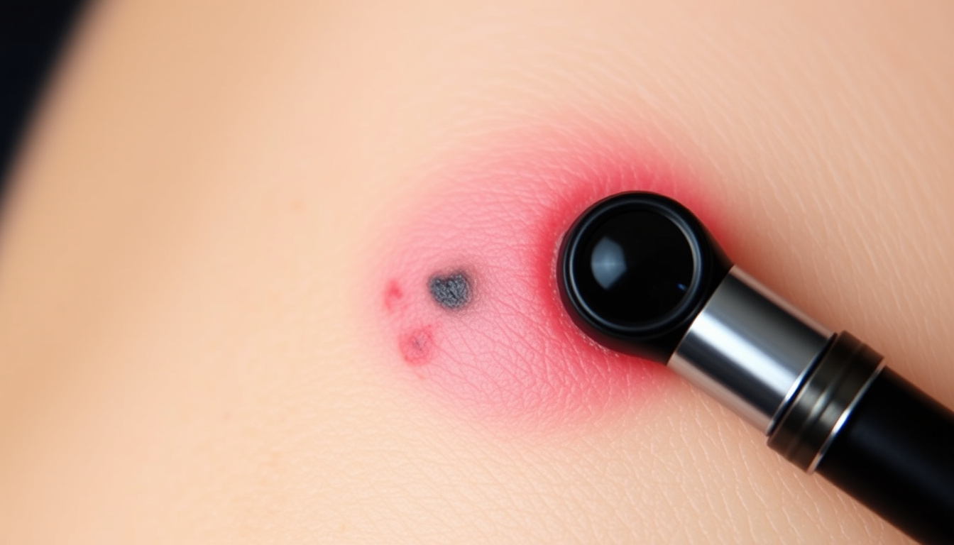
What Do Cancerous Moles Look Like? (With Pictures)
The single most important visual clue is a mole or spot that looks different from others on your body or that changes over time. Dermatology image archives show that cancerous moles often display asymmetry, irregular borders, multiple colors, and a diameter larger than about 6 mm—roughly the size of a pencil eraser. However, not all melanomas follow these rules, and some benign moles can appear concerning.
Visual Gallery
Browse medically reviewed pictures of cancerous moles, melanomas, and other skin cancers.
ABCDE Rule
Learn how to check moles for asymmetry, border irregularity, color variation, diameter, and evolution.
Types of Skin Cancer
Compare images of basal cell carcinoma, squamous cell carcinoma, and melanoma side-by-side.
When to See a Doctor
Find out which moles need professional evaluation and how to perform a skin self-exam.
- Early‑stage melanomas are often small and can mimic benign moles – the ABCDE rule is critical.
- Basal cell carcinoma often appears as a pearly or waxy bump, while squamous cell carcinoma may be a red, scaly patch.
- Melanoma can develop on areas not exposed to the sun, including palms, soles, and under nails.
- The most common location for melanoma in men is the back; in women, the legs.
- Over 90% of melanomas are curable when detected early.
- A mole that is new or changing in an adult over 30 should be evaluated regardless of ABCDE criteria.
| Feature | Benign Mole | Melanoma | Other Skin Cancers |
|---|---|---|---|
| Shape | Symmetrical | Asymmetrical | Often symmetrical (BCC/SCC may be irregular) |
| Border | Smooth, even | Irregular, ragged, notched | BCC: rolled border; SCC: scaly, crusty |
| Color | Single shade (tan, brown) | Multiple colors (black, brown, tan, blue, red) | BCC: pearly, pink; SCC: red, scaly |
| Diameter | Usually <6 mm | Often >6 mm | BCC/SCC can be variable |
| Evolution | Stable over time | Changes in size, shape, color, symptoms | BCC grows slowly; SCC may grow quickly |
How to Identify Melanoma Using the ABCDE Rule
The ABCDE rule is the most widely taught method for detecting melanoma. It stands for Asymmetry, Border irregularity, Color variation, Diameter, and Evolving. Developed by dermatologists, this mnemonic helps people recognize suspicious features that differ from normal moles.
What each letter means
- A – Asymmetry: One half of the mole does not match the other half.
- B – Border: Edges are uneven, ragged, blurred, or notched.
- C – Color: Multiple colors (brown, black, tan, red, white, blue) or uneven distribution.
- D – Diameter: Larger than about 6 mm, though some melanomas are smaller.
- E – Evolving: Any change in size, shape, color, elevation, or symptoms like itching or bleeding.
Compare the spot to all the other moles on your skin. A lesion that looks unlike the rest can be suspicious even if it doesn’t perfectly fit the ABCDE rule. This “ugly duckling” sign is a strong additional clue.
The ABCDE rule catches approximately 90% of melanomas. Exceptions exist, such as nodular melanoma, which is often symmetrical and uniformly colored. A changing mole or one that bleeds or fails to heal should be examined immediately.
What Are the Different Types of Skin Cancer and Their Pictures?
The three most common skin cancers are basal cell carcinoma (BCC), squamous cell carcinoma (SCC), and melanoma. Each has distinct visual features, though some may look similar. Below is a breakdown based on trusted medical image galleries.
Basal cell carcinoma
BCC often appears as a pearly or waxy bump, sometimes with visible blood vessels. It may also look like a flat, flesh‑colored scar-like patch. BCC rarely spreads but can destroy surrounding tissue if untreated. Skin Cancer Foundation pictures show these lesions commonly on sun‑exposed areas like the face and neck.
Squamous cell carcinoma
SCC often presents as a red, scaly, crusty patch or a firm, dome‑shaped growth that may bleed. It can develop from actinic keratoses, which are rough sun‑damaged spots. American Cancer Society image galleries illustrate SCC on the head, neck, hands, and arms.
Melanoma
Melanoma is the most dangerous form. It often appears as a new spot or a change in an existing mole. Cancer Research UK’s photos of abnormal moles highlight the classic ABCDE features. Some melanomas are amelanotic—lacking dark pigment—and may appear pink, red, or skin‑colored, making them harder to spot.
BCC and SCC are non‑melanoma skin cancers. They rarely metastasize, but an untreated SCC can spread. Melanoma is more aggressive and requires prompt surgical removal. The medical literature on PubMed emphasizes that early diagnosis dramatically improves prognosis.
Melanoma Pictures by Body Location: Legs, Face, Back
Melanomas can vary in appearance depending on their location on the body. Understanding these site‑specific patterns can improve self‑detection. According to epidemiological data, the back is the most common site in men, while the legs are most common in women.
Melanoma on legs
On the lower limbs, melanomas often appear as dark, irregular patches. Women frequently find them on the calves and thighs. NHS guidance on melanoma notes that any new or changing spot on the legs should be checked, especially if it has irregular borders or multiple colors.
Melanoma on face
Facial melanomas may be harder to detect because they can resemble age spots or freckles. Lentigo maligna, a type of melanoma in situ, appears as a slowly enlarging, flat, brown patch on sun‑damaged skin. Asymmetry and border irregularity are key clues.
Melanoma on back
The back is a common site for melanoma in men. Because it is hard to see, partner assistance or a mirror during self‑exams is useful. Lesions here often follow the ABCDE pattern and may be large (>6 mm) when discovered.
Early Stage Melanoma vs Benign Moles: Side-by-Side Comparison
Differentiating an early melanoma from a benign mole can be challenging. The most reliable approach combines the ABCDE rule, the ugly duckling sign, and attention to change over time. Below is a structured comparison based on visual features.
Images can raise awareness, but only a biopsy can confirm cancer. Many moles with concerning features are benign. Nodular melanoma and amelanotic melanoma may not follow the ABCDE rule at all. If you find a mole that is new, changing, bleeding, itchy, or crusting, see a dermatologist regardless of ABCDE.
- Benign moles: typically symmetrical, smooth‑bordered, one uniform color, stable size (often <6 mm), and round or oval.
- Early melanoma: often asymmetrical, irregular or notched borders, multiple colors (black, brown, tan, red, blue, white), diameter >6 mm, and evolving in size, shape, color, or symptoms.
- Key distinction: Benign moles rarely change, bleed, itch, or crust. Any such symptom warrants a professional check.
How Does a Mole Progress to Melanoma? (Timeline)
Melanoma development is not always linear, but a general progression from normal mole to invasive cancer is recognized. The following timeline describes typical stages, though not every mole follows this path.
- Normal mole: round, symmetrical, uniform color, stable size. Remains unchanged for years.
- Atypical (dysplastic) nevus: irregular border, mixed colors, larger than a normal mole. May appear over decades. Many do not progress further.
- Early melanoma (radial growth phase): asymmetry, uneven color, diameter >6 mm, evolving shape. This stage can last weeks to months.
- Invasive melanoma (vertical growth phase): lump, ulceration, bleeding, itching. High risk of spreading. This stage can develop over months to years.
The transition from a stable dysplastic nevus to melanoma is not inevitable. Regular monitoring helps capture changes early. The Cancer Research UK mole gallery provides examples of each stage.
What Is Certain and Uncertain When Using Pictures?
Medical images are powerful educational tools, but they have limits. Understanding what is well‑established versus what remains uncertain helps avoid false reassurance or unnecessary worry.
| Established information | What remains unclear |
|---|---|
| The ABCDE rule detects about 90% of melanomas when applied correctly. | Nodular melanomas can be symmetrical, uniformly colored, and small, making them missable by ABCDE. |
| BCC and SCC have distinguishing features (pearly bump vs. scaly patch). | Some BCCs mimic benign growths; some SCCs resemble warts or ulcers. |
| A biopsy is the only definitive diagnostic method. | No picture or AI app can replace a dermatologist’s examination. |
| New or changing moles in adults over 30 warrant evaluation. | The exact risk of a given atypical mole becoming melanoma is not precisely predictable. |
Why Are Pictures an Important Part of Skin Cancer Awareness?
Visual reference materials help bridge the gap between medical knowledge and public understanding. The ABCDE rule was developed by dermatologists specifically for laypeople. Image galleries from trusted sources such as the American Cancer Society, Skin Cancer Foundation, and NHS allow individuals to familiarize themselves with the appearances of normal moles versus suspicious lesions.
Australia, which has the highest melanoma rates globally, has long used public health campaigns featuring images of changing moles. In the UK and US, similar efforts have increased awareness. Regular skin self‑exams, combined with annual professional checks for high‑risk individuals (fair skin, many moles, family history), remain the standard recommendation.
Recent advances in AI‑based mole screening tools—including smartphone apps—are expanding access to initial assessments. However, these tools vary widely in accuracy and are not FDA‑approved for diagnosis. Caution is advised; any concerning result should be followed up by a dermatologist.
What Do Official Sources Say About Skin Cancer Images?
Authoritative health organizations consistently emphasize that pictures are for education, not self‑diagnosis. The following quotes reflect their guidance.
“Most melanomas come in the form of a new spot on the skin, not an existing mole.”
American Cancer Society, Skin Cancer Image Gallery
“The ABCDE rule should not replace a visit to a dermatologist – it’s a screening tool.”
Skin Cancer Foundation
“If you notice a mole that is different from others, or changes, itches, or bleeds, see your doctor.”
National Health Service (NHS)
“Basal cell carcinomas rarely spread, but squamous cell carcinomas can if untreated.”
Cancer Research UK
Summary: What Should You Take Away from This Guide?
The most reliable early warning signs of melanoma are a new or changing mole that is asymmetric, has irregular borders, shows multiple colors, and is larger than a pencil eraser. Benign moles are typically small, symmetric, even‑colored, and stable. For a deeper look at specific visual comparisons, see our benign vs. malignant mole comparison guide and our pictures of early melanoma and cancerous mole identification resource. Remember: no image can replace a clinical examination. If in doubt, see a doctor.
Frequently Asked Questions
Can a cancerous mole be completely flat?
Yes, early melanomas can be flat (in situ) and may not have a raised texture. Look for changes in color or border.
Do all cancerous moles itch?
Not all. Itching can be a sign, but many melanomas are asymptomatic. Bleeding, crusting, or scabbing are more concerning.
How often should I check my moles?
Monthly self‑exams are recommended for everyone. High‑risk individuals should have a yearly professional skin exam.
What does skin cancer look like on dark skin?
Melanoma on dark skin often appears on palms, soles, under nails, or in mucous membranes. It may be dark brown or black with irregular borders.
Are raised moles more likely to be cancerous?
Not necessarily. Many benign moles are raised. The shape, color, and border are more important than elevation.
Can I use an app to diagnose my mole?
Some apps use AI to analyze moles, but they are not FDA‑approved for diagnosis. Always see a doctor for confirmation.
Rebecca Haines is Managing Editor at DailyCity.co.uk, running the daily news list and covering UK and world stories with a city lens.